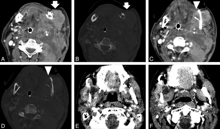
Fig 2.
Images depicting soft-tissue findings of recurrent tumor. Axial CT scan from a 62-year-old man with recurrent oral cancer shows a large mass (white arrows) adjacent to the left mandibular angle in soft-tissue (A) and bone (B) algorithms. Axial CT scan at a different level shows an area of lucent trabecular loss involving the left mandibular angle (white arrowheads) in soft-tissue (C) and bone (D) algorithms. Axial CT images in an 83-year-old patient with recurrent oral cancer show another finding seen in recurrent tumor, a cystic mass (curved arrows), at 2 different levels (E and F).

