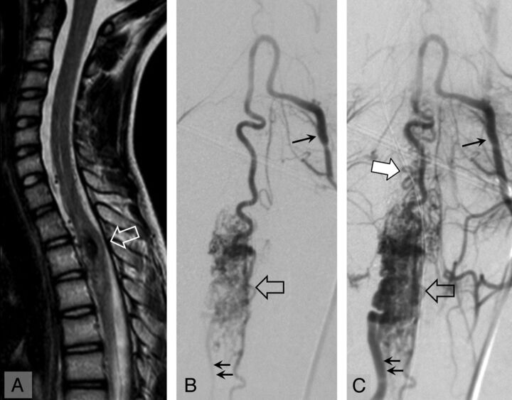
Fig 3.
Spinal cord MR imaging (A, sagittal T2 TSE) and angiography, anteroposterior view, early (B) and late (C) arterial phase in a 15-year-old girl with an upper thoracic spinal cord AVM at the time of recurrence of hematomyelia (29 months' follow-up). On MR imaging, the hematomyelia and AVM are located at T2 (open arrow). On angiography, the AVM (open arrow) is fed by a radiculomedullary artery (single arrow). The AVM is drained inferiorly by a radicular vein (double arrows) and superiorly by perimedullary veins (white arrow).

