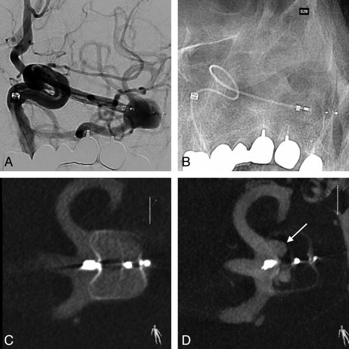
Fig 3.
A, DSA before detachment of a WEB positioned in a left MCA bifurcation aneurysm. It is impossible to depict any protrusion of the device in the parent artery because only the 3 markers are seen and the mesh is almost not visible. B, Corresponding unsubstracted image. C, VasoCT confirms correct positioning of the WEB without any protrusion. D, Three-month control VasoCT shows residual flow in the proximal compartment (arrow).

