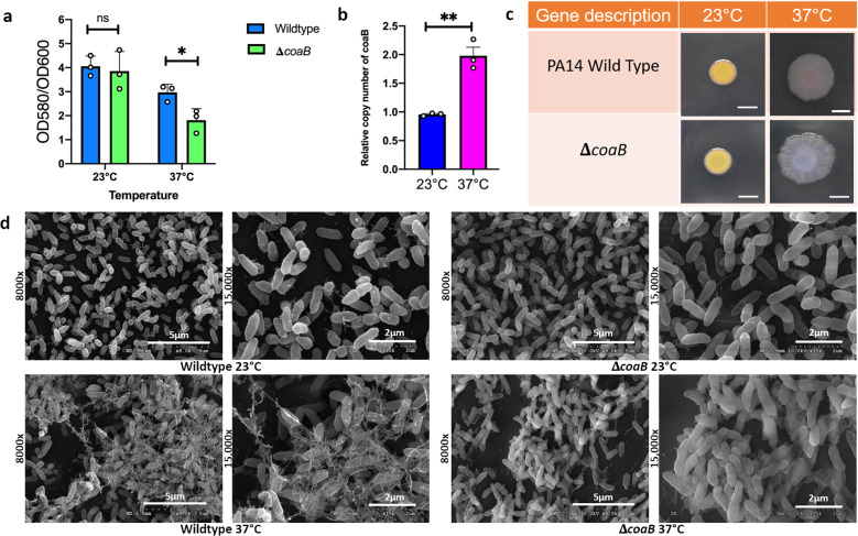
Fig. 4. The Pf1 filamentous phage coat B protein specifically contributes to P. aeruginosa PA14 biofilm EPS at human body temperature but not at room temperature.
a Crystal violet staining was performed to assess air-liquid biofilm formation in a 96-well microtiter plate at 48 h. Strains were grown in LB broth at appropriate temperatures. Bars represent the mean of three biological replicates performed on different days. The mean of each biological replicate was based on three technical replicates. Error bars represent the standard error of mean of the biological replicates. Unpaired t-test (two-tailed) was used to measure statistical significance. *p = 0.038, ns, p = 0.6071. b qPCR was performed on DNA extracted from biofilm grown for 48 h at 23 and 37 °C using primers specific for coaB and the housekeeping gene rplU using an established mechanical dissociation method42. Bars represent the mean of three biological replicates performed on different days. The mean of each biological replicate was based on three technical replicates. Error bars represent the standard error of mean of the biological replicates. Unpaired t-test (two-tailed) was used to measure statistical significance. **p = 0.0028. c Congo red binding assay. Extracellular matrix production by the wild type and mutant strain was evaluated on tryptone agar plates containing Congo Red and Coomassie brilliant blue G after incubation at 23 and 37 °C for 72 h. Representative images of the colony morphologies of WT PA14 and the ΔcoaB mutant are shown. Scale bar: 5 mm. d Comparison of the scanning electron microscopy images of Pseudomonas aeruginosa wild-type biofilms and ΔcoaB mutant biofilms grown for 48 h at 23 and 37 °C. Images show ×8000 and ×15,000 magnification and are representative of three independent experiments.

