Figure 2.
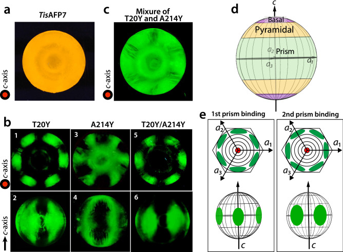
Ice plane specificity of wild-type TisAFP7 and its mutants, as determined by FIPA analysis. The concentration of protein solution was 0.007 mg/ml in all experiments. (a) Fluorescence image of a single ice-crystal hemisphere grown in a solution of wild-type TisAFP7 labeled with an orange fluorescent dye. The single ice crystal was mounted on the cold finger perpendicular to the basal plane. The c-axis direction of the ice crystal is shown in the figure as a circle. (b) Fluorescence from single ice-crystal hemispheres grown in solutions of TisAFP7 T20Y, A214Y, and T20Y/A214Y mutants labeled with a green fluorescent dye. Upper panels, the hemispheres were mounted in the same orientation as in (a); lower panels, the hemispheres were mounted with a primary prism plane perpendicular to the cold finger. The c-axis direction is shown as an arrow. (c) A single ice crystal grown in the solution containing equal concentrations of T20Y and A214Y mutants (0.0035 mg/ml of each). (d) Schematic illustration of ice planes mapped onto a hemispherical-shaped ice crystal. The polar regions correspond to the ice basal plane. The equator and middle latitude zones correspond to the prism and pyramidal planes, respectively. (e) Schematic illustrations of fluorescent patches corresponding to the primary and secondary prism planes on the ice hemisphere in known orientation. When the ice crystal is mounted with the basal plane perpendicular to the cold finger, the primary (1st) prism plane appears between the a1-, a2-, and a3-axes with a hexagonal symmetry, as shown by the upper images. The secondary (2nd) prism plane is situated on the region pierced with the a1-, a2-, and a3-axes. When the ice crystal is mounted with the primary prism plane perpendicular to the cold finger, three patches are observed on the equator of the hemisphere for the primary prism plane, whereas two patches are seen for the secondary prism plane.

