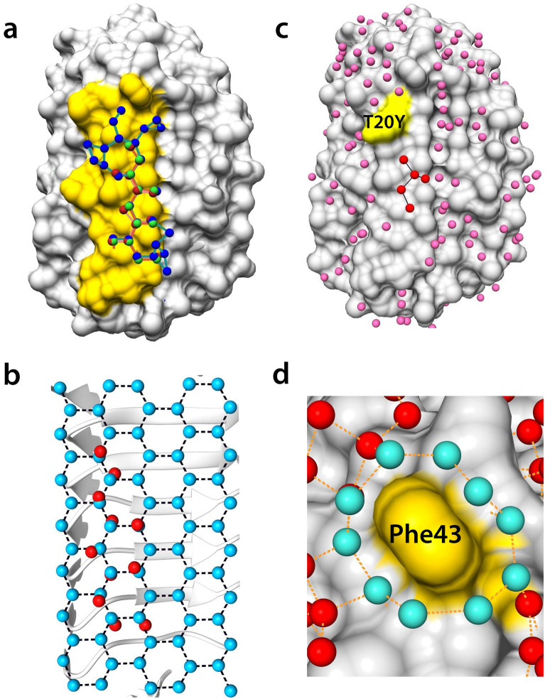
Figure 6.
Bound water network and superposition on the basal ice-plane water. (a) Surface structure of TisAFP7, with bound water molecules in the loop region of ice-binding site (IBS) colored in yellow. The bound water molecules in TisAFP7, TisAFP8, and TisAFP6 are drawn as spheres in red, blue, and green, respectively. Proximal water molecules, within 3.7 Å, are connected by solid lines. (b) Eleven bound water molecules at the IBS loop of TisAFP7 superposed on the basal plane water molecules of an ice crystal with root-mean–squared deviation of 0.77 Å. (c) Crystal structure of the T20Y mutant with bound water molecules on its surface. The altered site (Tyr20) is indicated in yellow. IBS water molecules are denoted in red and other bound water molecules in pink. (d) Magnified view of Phe43 surrounded by a water ring composed of 10 water molecules drawn in cyan. Each water molecule located within 3.5 Å of each other is connected by a dashed line.

