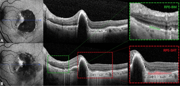
Figure 2.
Measurement of repair tissue reflectivity following retinal pigment epithelial (RPE) tear by means of spectral-domain optical coherence tomography (SD-OCT). (A) and (B) show blue-light autofluorescence and SD-OCT scans of a patient at baseline and at 12 months of follow-up, respectively. Red dashed box highlights the formation of the RPE-subretinal hyperreflective tissue complex that was quantitatively measured after manual delineation. RPE-Bruch’s membrane complex, used as internal reference, is presented in the green dashed box.

