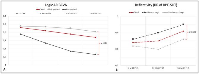
Figure 3.
Variation of the best-corrected visual acuity (BCVA) and of the relative reflectivity (RR) of the repair tissue over the follow-up. (A) shows the visual acuity changes occurring in time; BCVA appears to deteriorate remarkably in patients that did not have signs of repair tissue formation (p = 0.04). The reflectivity of the repair tissue (B) increases in time during the follow-up particularly in patients with hemorrhagic tears at baseline (p = 0.001).

