Fig. 3.
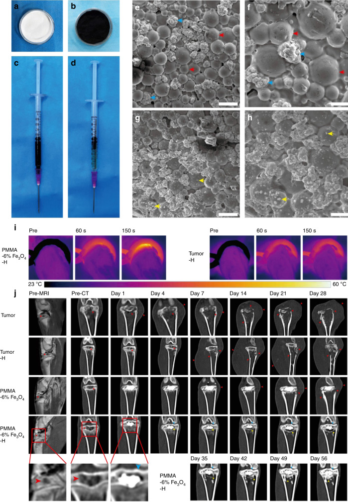
PMMA-Fe3O4 for magnetic ablation of bone tumors and bone repair. a PMMA powder, b Fe3O4 nanoparticles, c MMA monomer, and d injectable PMMA-6% Fe3O4. e Low-magnification SEM image of polymerized PMMA. The scale bar is 50 μm. f High-magnification SEM image of polymerized PMMA. The scale bar is 20 μm. g Low-magnification SEM image of polymerized PMMA-6% Fe3O4. The scale bar is 50 μm. h High-magnification SEM image of polymerized PMMA-6% Fe2O3. The scale bar is 20 μm. i Thermal images of rabbit legs in the PMMA-6% Fe3O4–H group and Tumor-H group. j Enhanced MRI images and coronal reconstructed CT images at each follow-up time point (red arrow: bone destruction and swelling of soft tissue, blue arrow: cortical bone of upper tibial plateau, yellow arrow: area of bone resorption and new bone formation). Reprinted with permission from ref. 113 © 2019, Ivyspring International Publisher

