
Figure 7.
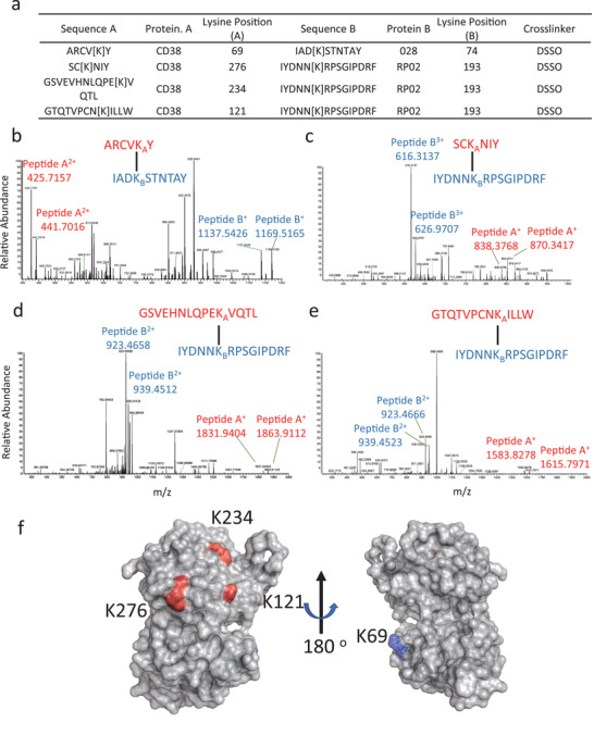
Identification of RP02 and 028 binding site on the hCD38‐ECD. a) hCD38‐ECD was crosslinked with RP02 or 028 scFv using DSSO. Crosslinked peptides were identified by high‐resolution MS analyses. b–e) Secondary MS spectra identified crosslinked peptides ARCVKY‐IADKSTNTAY, SCKNIY‐IYDNNKRPSGIPDRF, GSVEHNLQPEKVQTL‐ IYDNNKRPSGIPDRF, and GTQTVPCNKILLW‐ IYDNNKRPSGIPDRF. Tertiary MS data are shown in Figures S6–S9 (Supporting Information). The MS data were deposited in the proteomics identifications (PRIDE) database with accession number PXD019713. f) Location of RP02 and 028 binding sites on hCD38‐ECD. The lysine crosslinked with 028 (K69) is shown in blue and the lysine crosslinked with RP02 (K121, K234, and K276) is shown in red. The structure of hCD38‐ECD (PDB 1yh3) is displayed by PyMOL.