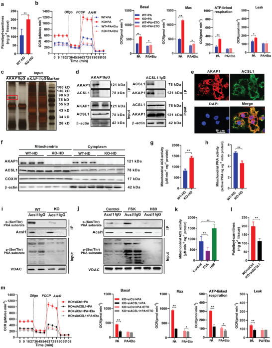
Figure 5.

AKAP1 decreases fatty acid β‐oxidation by inhibiting ACSL1 activity. a) Palmitoyl carnitine level was measured by LC–MS/MS in BAT of WT and AKAP1−/− HFD mice (n = 6 mice per group). b) Fatty acid oxidation (FAO) profiles were determined by a Seahorse extracellular flux analyzer in differentiated WT and AKAP1−/− brown adipocytes after exposure to 500 µM palmitate for 24 h. Cells were treated sequentially with Oligo (3 µM), the chemical uncoupler FCCP (1 µM), R( 4 µM), and AA(4 µM) in the presence of palmitate (PA). Etomoxir (Eto, 40 µM) was used to inhibit FAO and to confirm the assay specificity. Data were from three independent experiments. c) IP assay using AKAP1 antibody or immunoglobulin G (IgG). Samples were run on Bis‐Tris Gel and then stained with Fast Silver Stain Kit. IgG, negative control antibody. d) Co‐IP assay showing interaction between AKAP1 and ACSL1 using AKAP1 antibody (left) or ACSL1 antibody (right). e) Representative images of immunofluorescence for ACSL1 (green), AKAP1–Flag (red), and nuclei (blue) in brown adipocytes infected with AAVDJ–AKAP1–Flag. Scale bar, 10 µm. f) Western blotting analysis of AKAP1 and ACSL1 expression in mitochondria or cytoplasm of BAT from WT and AKAP1−/− mice on HFD (n = 3 mice per group). Mitochondrial cytochrome c oxidase subunit IV (COXIV) and β‐actin were used as loading controls. g) Mitochondrial ACS activity in BAT from WT and AKAP1−/− mice on HFD (n = 6 mice per group). h) Mitochondrial protein kinase A (PKA) activity in BAT from WT and AKAP1−/− mice on HFD (n = 6 mice per group). i) The lysate of BAT mitochondria was immunoprecipitated using ACSL1 antibody and blotted with an anti‐phospho‐(Ser/Thr) PKA substrate antibody in WT and AKAP1−/− mice on HFD. Voltage‐dependent anion channels (VDAC) was used as a loading control. j) The lysate of BAT mitochondria was immunoprecipitated using ACSL1 antibody and blotted with anti‐phospho‐(Ser/Thr) PKA substrate antibody in HFD mice treated with PKA activator forskolin (FSK) or PKA inhibitor H89. 50 µL of FSK (80 µM) or H89 (10 µM) was injected into interscapular BAT. BAT samples were collected 2 h after injection. VDAC was used as a loading control. k) Mitochondrial ACS activity in BAT from mice treated with PKA activator (FSK, 80 µM) or PKA inhibitor (H89, 10 µM). n = 6 mice per group. l) Palmitoyl carnitine levels were measured by LC–MS/MS in siRNA‐injected BAT of AKAP1−/− mice (n = 6 mice per group). siACSL1: siRNA against ACSL1; siCtrl: control siRNA. m) Fatty acid oxidation (FAO) profiles in AKAP1−/− brown adipocytes with siRNA transfection were determined by a Seahorse extracellular flux analyzer. Data were from three independent experiments. Data were expressed as mean ± SEM. Student's t‐test was used in (a), (b), (g), (h), (l), and (m). One‐way ANOVA with Bonferroni's post hoc test was used in (k). *p < 0.05, **p < 0.01.
