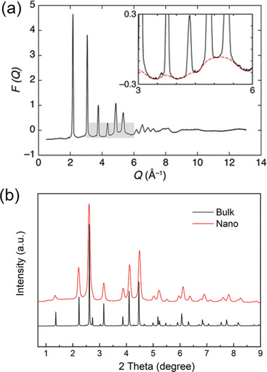
Figure 1.

a) Illustration of diffraction and diffuse scattering signals spreading over Q‐space. The F(Q) presented here was normalized from the diffraction of AgBr. The inset emphasizes the diffuse signals associated with structural disorders. Reproduced with permission.[ 19 ] Copyright 2011, Royal Society of Chemistry. b) Comparison of synchrotron XRD patterns of MnFe2O4 bulk and nanoparticles collected at 11‐ID‐C beamline (λ = 0.1173 Å).
