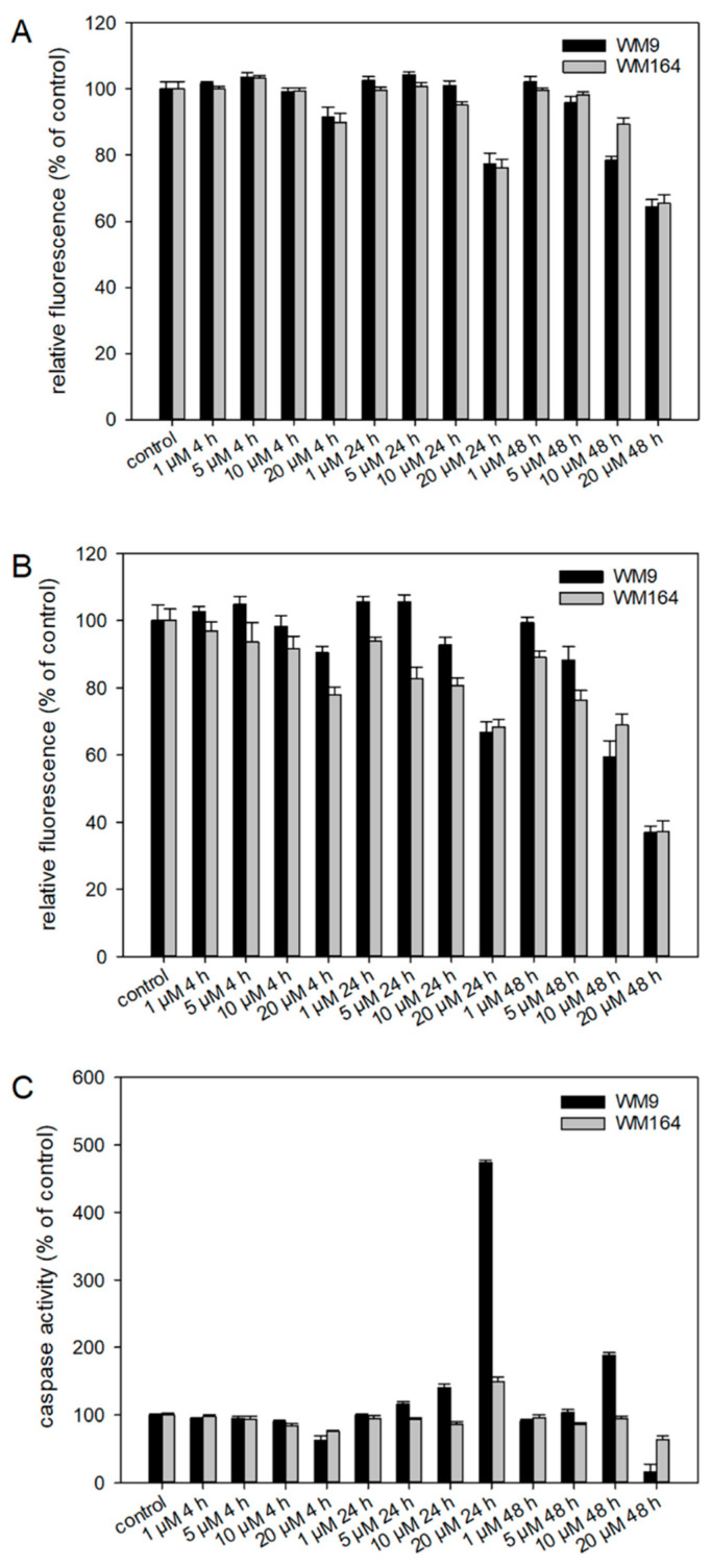
Figure 6.
Results of the ApoToxGlo™ Triplex Assay. WM9 and WM164 cells were treated with 1 µM, 5 µM, 10 µM, and 20 µM of 5 for 4 h, 24 h, or 48 h (n = 6, mean ± sem). (A) Viability of the cells measured as relative fluorescence of control cells. (B) Cytotoxicity of 5 towards the cells measured as relative fluorescence of control cells. (C) Activity of caspases 3 and 7 indicative for apoptosis induction. Staurosporine (25 µM) served as positive control (apoptosis increase in WM9 cells after 24 h: 1193.9 % and after 48 h: 297.4%; in WM164 cells after 24 h: 989.1% and after 48 h: 362.6%).

