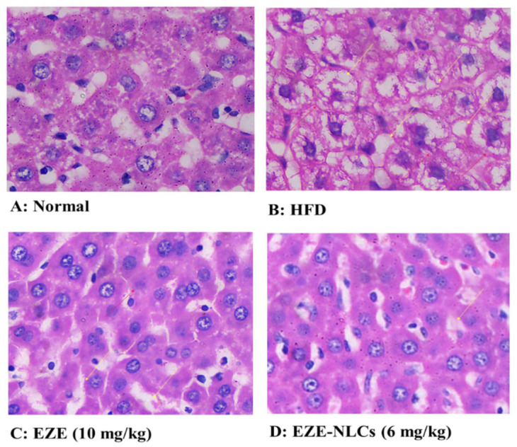
Figure 8.

Effect of EZE-NLCs on histopathological changes (10×). (A) shows the normal liver section exhibits regular liver architecture with normal polygonal hepatic cells; (B) HFD shows abnormal architectural organization of hepatic tissue, dilatation, enlargement of sinusoids, vascular congestion, vacuolization, and Kupffer cell infiltration; (C) and (D) shows improvements of hepatocyte structures, Kupffer cells and with without vacuoles was attained. It is clear that EZE-NLCs remarkably reduced the histological changes when compared to that observed in HFD fed rats and the EZE treated rats.
