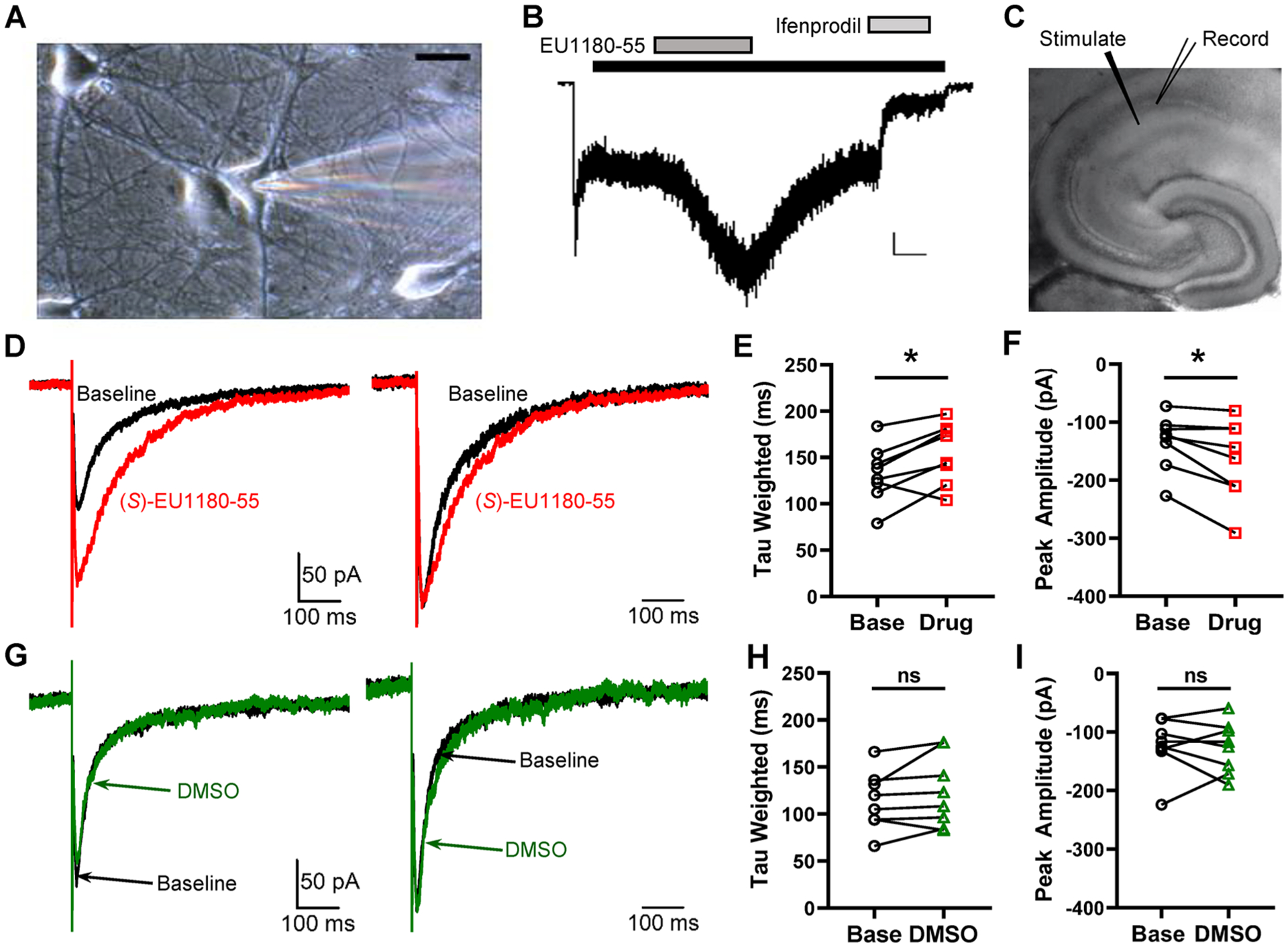
Figure 4.

Effect of EU1180–55 on NMDARs in hippocampal neurons. (A) Representative photomicrograph of a neuron maintained 12 days in culture (calibration 20 μm). The patch electrode can be seen on the right side. (B) Typical current response to 100 μM NMDA and 3 μM glycine (black bar). After the current relaxed to a steady-state level, EU1180–55 (10 μM, dark gray bar) was coapplied with NMDA and glycine, which increased the current response to NMDA in the presence of bicuculline (10 μM), CNQX (10 μM), and TTX (0.5 μM) to 200% ± 10% of control (p = 0.001, paired t test, n = 5). After wash out of EU1180–55, 3 μM ifenprodil, the GluN2B-selective inhibitor (light gray bar), was applied. Ifenprodil inhibited the current to 30% ± 10% of control (p = 0.005, paired t test, n = 5). The calibration bar is 50 pA, 20 s. (C) NMDAR-mediated component of evoked EPSCs recorded at −30 mV in the presence of 0.2 mM Mg2+ from 300 μm hippocampal slices from P7–14 mice. (D) Left panel, representative evoked NMDAR-mediated EPSC before (black) and after (red) the addition of 20 μM (S)-EU1180–55. Right panel, normalized evoked NMDAR-mediated EPSC before (black) and after (red) the addition of 20 μM (S)-EU1180–55. (E, F) Effect of (S)-EU1180–55 on weighted tau and response amplitude (n = 8, red). (G–I) Similar data as in panels D–F but with DMSO vehicle (n = 8, green). See Supplemental Table S5 for summary of fitted parameters. *p < 0.025, paired t test.
