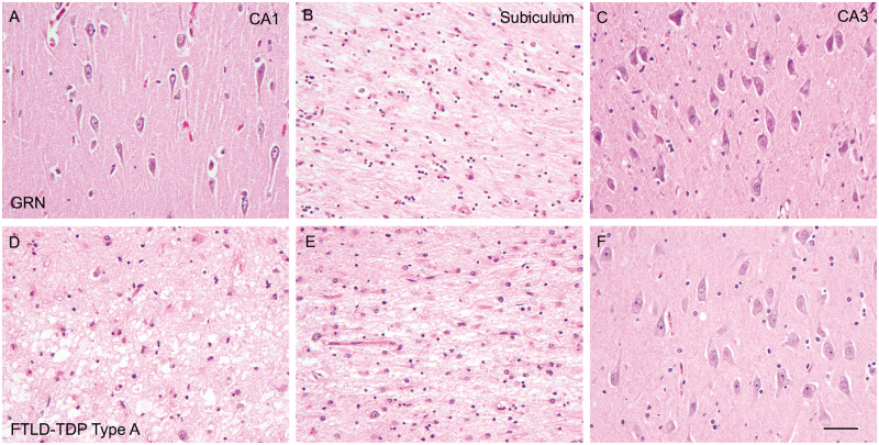
FIGURE 2.
Hematoxylin and eosin stains reveal unsynchronized neuronal loss in the CA1 region and subiculum of brains with GRN mutations. Neuronal loss is mostly mild in the CA1 region (A), severe in the subiculum (B), and minimal in the CA3 region (C) of FTLD-TDP type A with GRN mutation (GRN+/−) brains. However, neuronal loss is severe in both the CA1 region (D) and subiculum (E), and minimal in the CA3 region (F) of FTLD-TDP type A without GRN mutation brains (FTLD-TDP type A). Bar: 50 µm.

