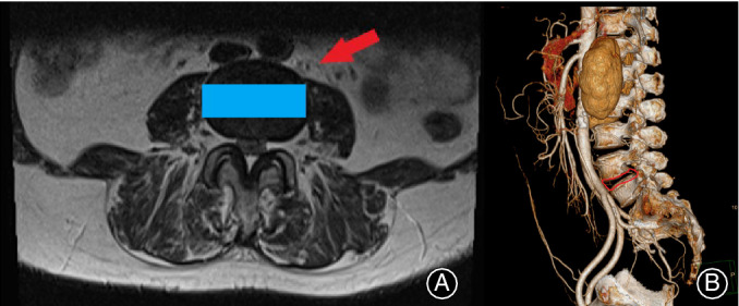
Figure 2.

(A) Arrow indicates the route to the intervertebral disc and the rectangle shows the position where the cage is inserted. Cross‐section of the MRI determines if it is suitable to perform oblique lumbar interbody fusion (OLIF). (B) CTA is used to find whether there is abnormal vascular malformation to decrease the incidence of vascular injury.
