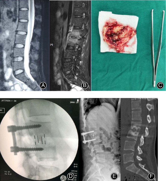
Figure 3.

The preoperative sagittal T2‐weighted MRI (A, B) of a 31‐year‐old female patient showed the disruption of intervertebral space 1 week and 2 months after the onset of low back pain, respectively. (C) The disrupted intervertebral disc. The lateral (D–F) radiographs and CT showed oblique lumbar interbody fusion at L2–3 levels intraoperatively, 3 and 6 month postoperatively. Bony fusion was achieved.
