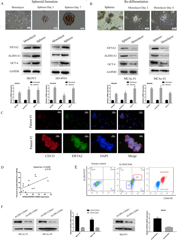
Fig. 2.
Upregulated EIF5A2 expression in OCSC-like cells. a Protein expression of EIF5A2, OCT-4, and ALDH1A1 of parental SKOV3, HO-8910 cells (monolayer), day 7 spheroid cells derived from SKOV3, HO-8910 cells growing in serum-free medium with growth factors (spheres), measured by western-blot. b Protein expression of EIF5A2, OCT-4, and ALDH1A1 of MCAs from two EOC ascites (spheres), adherent cells after re-differentiation growing in serum-complete medium (monolayer). c Immunofluorescent staining of CD133 (red), EIF5A2 (green), and their co-localization (yellow) in EOC malignant cell from ascites. d Correlation analysis demonstrating that overexpression of EIF5A2 was positively correlated with percent of ALDH+CD44+ subpopulation cells in MCAs from EOC patients. (r = 0.6268, p < 0.05). e Isolation of ALDH+CD44+ and ALDH−CD44− subpopulaiton cells by FACS. (Left) Isotype control. (Middle) The top 5% cells showing the highest staining for ALDH+CD44+ or bottom 5% with minimal staining for ALDH-CD44- cells were collected. (Right) Purity of ALDH and CD44 was confirmed via post sorting using flow cytometry. f Relative EIF5A2 mRNA and protein expression of ALDH+CD44+ and ALDH-CD44- subpopulation cells in MCAs #5, MCAs #9, and SKOV3 cells via qPCR and immunoblotting, respectively. All the data represent the means ± SD; *p < 0.05, **p < 0.01, and ***p < 0.001

