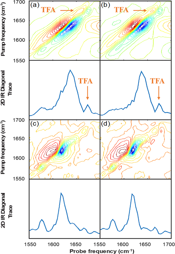
Figure 3.
2D IR spectra of (a) G24A25 amylin in depleted serum with 0.03% TFA added as an internal standard, (b) depleted human serum with 0.03% TFA added as an internal standard, and (c) G24A25 amylin in depleted human serum with the internal standard TFA signal used to scale the subtraction of the depleted serum background and, for comparison, (d) the same spectral subtraction where the TFA signal was not used.

