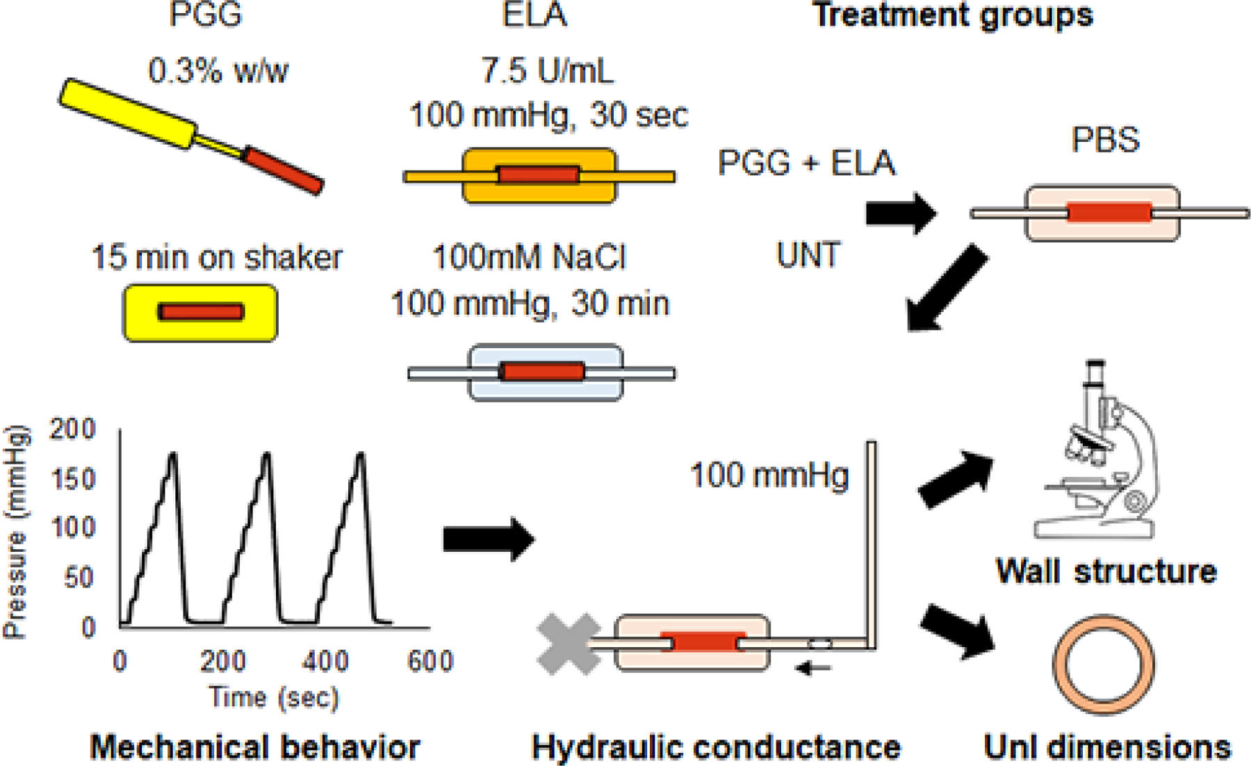
Fig. 1.

Diagram of the treatment groups and experimental procedures. Mouse carotid arteries were treated with PGG, ELA, PGG+ELA, or UNT. They were then mounted in a pressure myograph and cyclically inflated for three cycles under in vivo axial stretch. Next, a static pressure of 100 mmHg was applied and the movement of a bubble was recorded to measure fluid transport across the arterial wall. The arteries were then removed from the myograph. Small rings were cut to measure unloaded dimensions after treatment and testing and the remaining arterial tissue was processed for imaging the wall structure.
