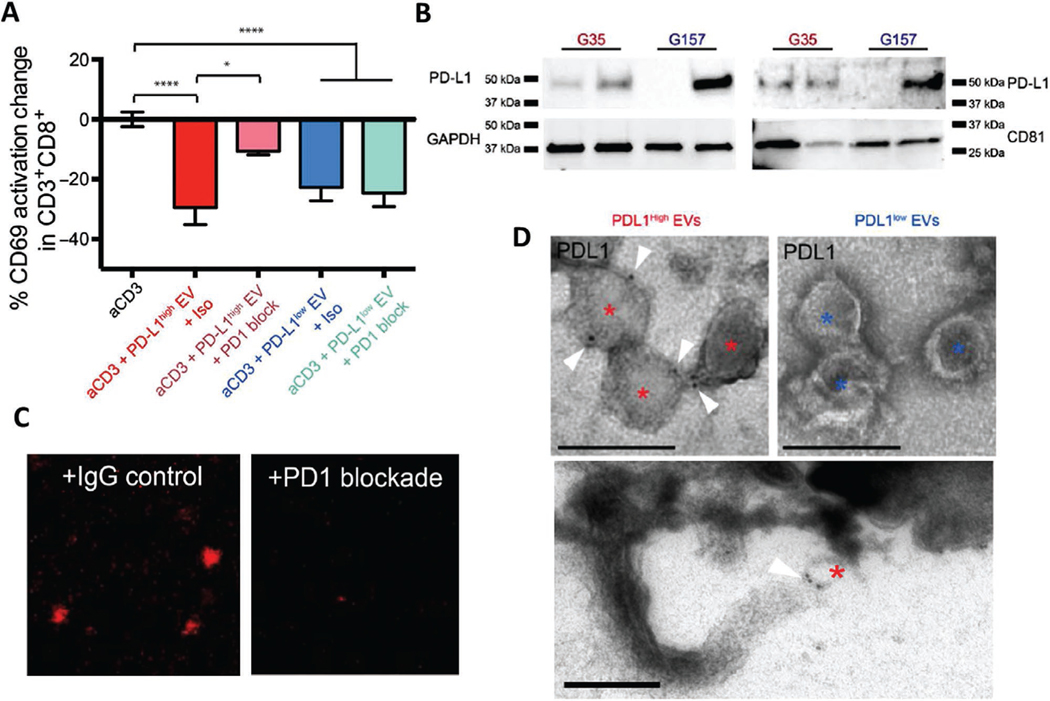
Figure 1.
PD-L1 carried on extracellular vesicles inhibits T cell activation via direct binding to PD-1. A) PD1 blockade prevents the inhibition of PD-L1high GSC EVs on PBMCs. Percent change in CD69 expression CD8+ T cells is shown as a measure of T cell activation. PD1 blocking antibody or isotype control was added at day 0 and CD69 levels were determined 48 h later (n = 7 PBMC donors, means ± SD). B) IFNγ-mediated increase of PD-L1 expression levels in PD-L1high and PD-L1low GSCs as shown by Western blots. C) Representative confocal microscopy images showing that fluorescently labeled EVs isolated from PD-L1high GSCs can bind to wells coated with recombinant PD1 (left panel). EV binding is displaced by co-incubation with an anti-PD1 antibody (right panel), indicating specificity of binding. D) Immuno-electron microscope images of PD-L1 immunolabeled PD-L1high and PD-L1low glioblastoma EVs are shown. Asterisks mark each EV, and the white triangle indicates gold-immunolabeled PD-L1, scale bar = 200 nm. Lower panel shows a PD-L1-labelled EV apparently ligated to a process at the lymphocyte surface. Reproduced under the terms and conditions of the Creative Commons Attribution, Non Commercial Licence 4.0.[19]

