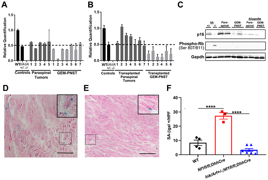
Figure 5. Ink4a/Arf mice show loss of heterozygosity and reduced senescence.

A, B. Q-PCR analysis of Ink4a/Arf shows rare LOH in solid tumors A. One GEM-PNST showed LOH; error bars show standard deviation. B. Of the grafts that enlarged after transplantation, half of GEM-PNST, but no paraspinal tumors, showed LOH; error bars show standard deviation. C. Western blot of p16 protein shows reduction in protein expression in some solid tumors and all nerve grafts. At left, controls are lysates of lung from Ink4a/Arf+/− and tumor from Ink4a/Arf−/− mice. D, E. SA-β-gal-positive stained (blue) cells in DRG-associated nerve; pink is counterstain. Inserts show higher magnification micrographs. D. Nf1fl/fl;DhhCre. E. Ink4a/Arf+/−;Nf1fl/fl;DhhCre. F. Quantification of cells with positive beta-galactosidase staining/high powered field averaged over 10 hpf. Each mark represents a separate mouse. Tukey’s multiple comparisons test, p<0.0001=****.
