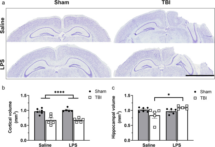
Fig. 12.
Volumetric analyses of intact brain tissue at 4 days post-LPS (i.e., 8 days post-surgery). Brain tissue was stained with Cresyl violet solution (Sham + saline, Sham + LPS, TBI + saline, and TBI + LPS representative images are shown in box. a) and ipsilateral hemisphere observations were normalized to contralateral hemisphere. A significant reduction in cortical tissue volume was observed in TBI groups compared to sham groups (b). However, the hippocampal volume (c) was significantly increased in TBI + LPS group compared to the TBI + saline group. *p < 0.05 from post-hoc; ****p < 0.001 main effect of injury from two-way ANOVA. n = 5–6/group. Scale bar = 2000 μm

