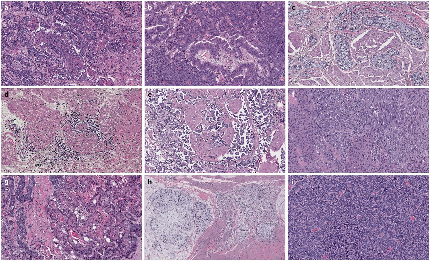
Fig. 3 |. Variant histology of urothelial carcinoma.

Tumour heterogeneity is most pronounced at the morphological level when comparing urothelial carcinomas with variant histology. The morphological spectrum of urothelial carcinoma includes divergent differentiation, such as squamous differentiation (part a) and glandular differentiation (part b). Variant histologies of urothelial carcinoma include nested variant (part c), plasmacytoid variant (part d), micropapillary variant (part e) and sarcomatoid variant (part f). Primary tumours of non-urothelial histology can also develop in the bladder, including squamous cell carcinoma (part g), mucinous adenocarcinoma with signet ring cells (part h) and small-cell or neuroendocrine carcinoma (part i). Haematoxylin and eosin staining in all images, magnification ×50.
