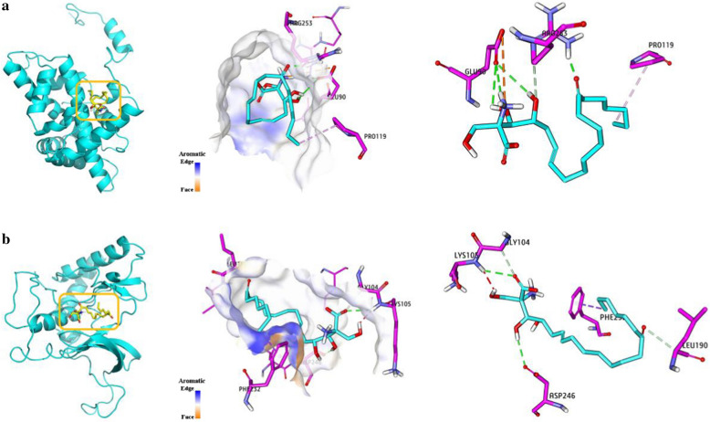
Fig. 7.
Molecular docking model of myriocin-NFYA/RIOK2. a and b represent the models of myriocin-NFYA and myriocin-RIOK2, respectively. From left to right, ribbons, surfaces and detailed binding diagrams are shown. Left: rectangular boxes represent internal docking areas. Middle: detailed internal docking areas (the surface color of proteins is classified by aromaticity). Right: detailed the bonding of myriocin-NFYA/RIOK2. GLU90, ARG253, PRO119, LYS105, ASP246, GLY104, LEU190, PHE232, PRO195 and ILE235 are amino acid residues. Hydrogen bonds are shown in green dashed lines. Electrostatic are shown in orange dashed line. Hydrophobics are shown in pink dashed line

