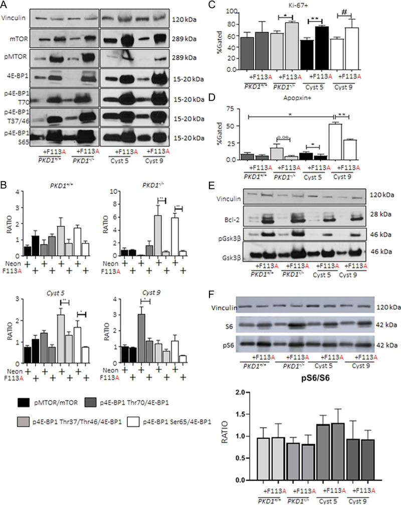
Figure 4.

4E-BP1F113Ain vitro promotes proliferation and retards apoptosis. PKD1+/+, PKD1−/− and Human Cyst 5 and 9 were transduced with either control neon-expressing, or HA-tagged 4E-BP1F113A constructs as described. (A) Loading control vinculin, total and phosphorylated mTOR and total and phosphorylated 4E-BP1 (Thr70, Thr37/Thr46 and Ser65) were detected and quantified (B) as the ratio of densitometry units for phosphorylated to total substrate abundance. (C) Proliferation marker (Ki-67), and (D) apoptosis marker (Apopxin) were assayed by flow cytometry. (E) Immunoblotting of anti-apoptotic proteins; Bcl-2, GSK3β (total, and phospho) were augmented in 4E-BP1F113A-transduced cell lines. (F) Increase in both phospho and total S6K. Representative immunoblots reflect a minimum of six independent protein isolations from successive passages. Multiple comparisons were made using two-way ANOVA. Values are expressed as the mean ± SEM. *P < 0.05, **P < 0.01, ***P < 0.001, #P < 0.0001.
