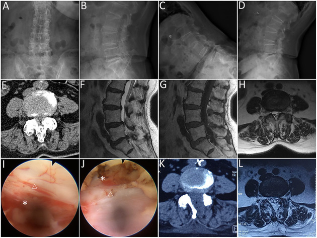
Figure 2.

Lumbar endoscopic unilateral laminotomy bilateral decompression (LE-ULBD) performed on a 77-year-old female patient diagnosed with L4-L5 lumbar spinal stenosis with degenerative spondylolisthesis. (A,B) preoperative anteroposterior and lateral plain radiographs; (C,D) preoperative flexion and extension radiographs; (E) preoperative computed tomography (CT) scans; (F–H) preoperative magnetic resonance imaging (MRI) scans; (I,J) medial facetectomy was performed to decompress the lateral recess and ensure adequate decompression of the traversing nerve root; (K) postoperative CT scans; (L) postoperative MRI scans. Snowflake, nerve root, triangle, dural sac. * is used to tell the readers where is the nerve root.
