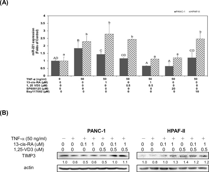
Fig 5. 13-cis RA and 1,25-VD3 induced the expression of TIMP3 through a suppression of miR-221 in TNF-α-stimulated PDAC cells.
(A) Human PDAC cells were treated with TNF-α in 10% FBS DMEM in the presence or absence of 13-cis RA, 1, 25-VD3, SP600125 or Bay117082 for 24hr. The effect of TNF-α on the expression of miR 221 was measured by using qPCR assay in the Materials and Methods section. Statistical significance is expressed as the mean ± SD (standard deviation) of two independent experiments. Different upper-case letters (A, B, C, D) represent statistically significant differences among subgroups in PANC-1 cells (P<0.05). Different lower-case letters (a, b) represent statistically significant differences among subgroups in HPAF-II cells (P<0.05). (B) Human PDAC cells were treated with TNF-α in 10% FBS DMEM in the presence or absence of 13-cis RA and 1, 25-VD3 for 24hr. Western blot analysis of cytoplasmic proteins was performed using monoclonal antibodies against TIMP-3 and actin, as described under Materials and Methods. Band intensities represent the amounts of TIMP-3 in the cytoplasm of human PDAC cells.

