
Figure 2:
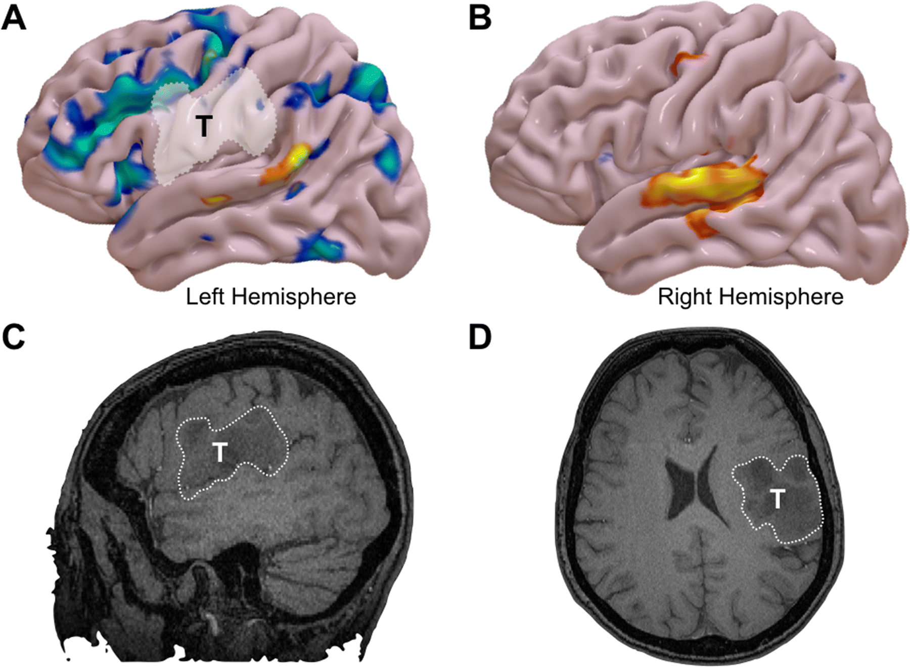
Reconstructive functional MRI (fMRI) results from a word-generation task in a bilingual patient with a WHO Grade III astrocytoma of the left frontoparietal operculum. A) The results for the patient’s native language is shown in blue and secondary language in orange. The native language shows an expected pattern of language activation in the left hemisphere, including both frontal and posterior language areas. B) fMRI representation of the secondary language in the right hemisphere. Non-contrast enhancing T1-weighted imaging showing the tumor in C) Left-sided sagittal cut and D) Axial cut on the left hemisphere.