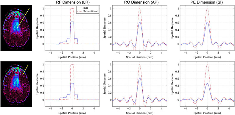
FIGURE 9.
SRFs for SER and conventional gSlider obtained from (top row) a voxel where the SNR improvement associated with SER was approximately 3 and (bottom row) a voxel where the SNR improvement associated with SER was approximately 5. The voxel positions are indicated as shown in the images on the left. The SRFs in this case are three-dimensional functions, which are hard to display. For easier visualization, we have shown one-dimensional plots passing through the peak of the SRF along different orientations. Specifically, we show SRF plots along the RF-encoding dimension (left-right anatomically), the readout encoding dimension (anterior-posterior anatomically), and the phase encoding dimension (superior-inferior anatomically)

