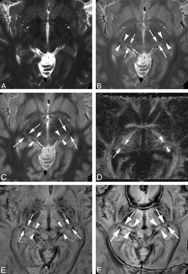
Fig 1.
MGN and LGN in a healthy 31-year-old female volunteer. T2-weighted (A), PD (B), STIR (C), DTI (D), original PADRE (1 mm) (E), and reconstructed PADRE (3 mm) images (F). A, Neither side of the MGN or LGN is identified (visualization score = 0 by both observers). B, The right MGN is poorly visible. Its boundary is fuzzy (short arrows) (visualization score = 1 by both observers). The left MGN is mostly visible; its boundary is slightly fuzzy (long arrows) (visualization score = 2 by both observers). Both sides of the LGN are mostly visible; the boundary is slightly fuzzy (arrowheads) (visualization score = 2 by both observers). C, One observer assigned a visualization score of 1, the other of 2, to the right MGN (short arrows). The left MGN is mostly visible with a slightly fuzzy boundary (arrowheads) (visualization score = 2 by both observers). Both sides of the LGN are mostly visible with a slightly fuzzy border (long arrows) (visualization score = 2 by both observers). D, Neither side of the MGN is identified (visualization score = 0 by both observers). The right LGN is mostly visible with a slightly fuzzy border (arrows) (visualization score = 2 by both observers). The left LGN is poorly visible; its border is fuzzy (arrowheads) (visualization score = 1 by both observers). E, Both sides of the MGN (arrowheads) and LGN (arrows) are well-identified and clearly differentiated from lateral and medial neighboring structures (visualization score = 3 by both observers). F, Although the boundaries of the LGN and MGN are slightly obscure on reconstructed PADRE compared with the original PADRE, both sides of the MGN (arrowheads) and LGN (arrows) are well-identified and clearly differentiated from lateral and medial neighboring structures (visualization score = 3 by both observers).

