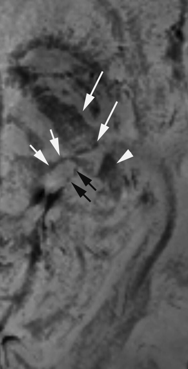
Fig 2.

A magnified PADRE image of the left MGN and LGN in a healthy 28-year-old female volunteer. The left LGN is surrounded by the cerebral peduncle (long arrows) anteriorly and the origin of the optic radiation (arrowhead) posteriorly. The MGN is surrounded by the inferior (arrows) and superior quadrigeminal brachium (black arrows). The MGN and LGN are distinguished from surrounding structures with low signal intensity.
