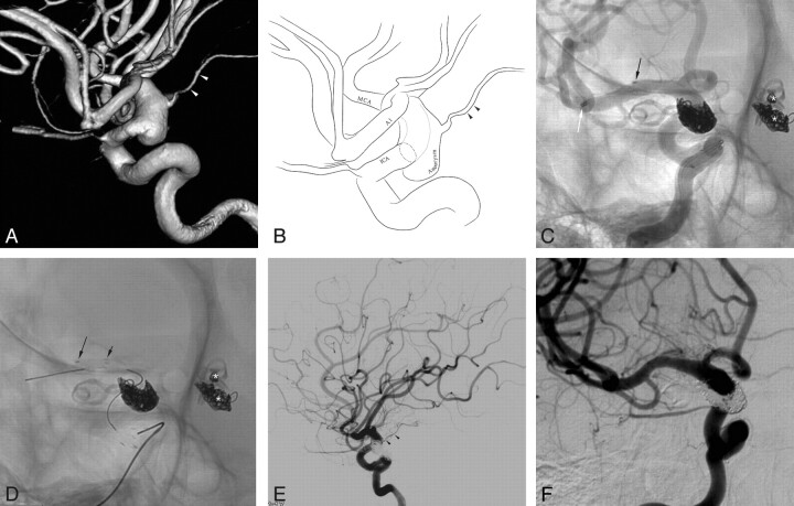
Fig 3.
A 30-year-old woman with a postclip recurrent aneurysm in the left posterior communicating artery, left anterior choroidal artery, and right anterior choroidal artery. The 3D reconstruction image (A) and its illustration (B) reveal a fusiform aneurysm of the right distal internal carotid artery with incorporation of the right anterior choroidal artery. C, Stent-assisted coil embolization was performed after deployment of the first Enterprise stent and re-advancement of the Prowler Select Plus microcatheter. White and black arrows indicate distal markers of the re-advanced Prowler Select Plus microcatheter and the deployed Enterprise stent, respectively. Asterisks indicate the coil-embolized left posterior communicating artery and anterior choroidal artery aneurysms. D, A second Enterprise stent was deployed after completion of coil embolization. The black arrows indicate distal markers of 2 overlapping Enterprise stents. E, Final control angiogram reveals near-complete occlusion of the aneurysm sac and preservation of the right internal carotid artery and the anterior choroidal artery (arrowheads). F, A 12-month follow-up angiogram in a working projection shows stable aneurysm occlusion.

