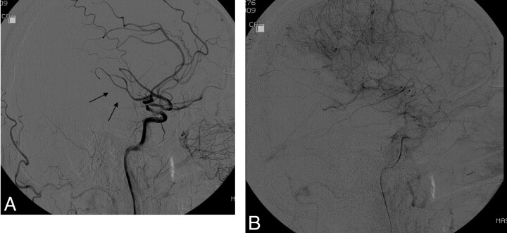
Fig 1.
A, Lateral DSA following acute stroke intervention shows delay in flow in the posterior division of the MCA (arrows) compared with the anterior cerebral artery branches. B, Late arterial phase shows opacification of nearly all of the MCA. Because this represents more than two-thirds of the MCA (and thus not TICI 2a) but not “complete filling” (and thus not TICI 2b), it cannot be categorized by using the original TICI classifications.

