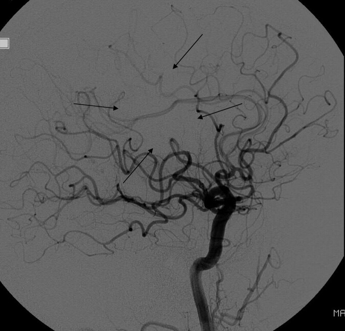
Fig 2.
Lateral DSA following acute stroke intervention shows a normal rate of antegrade flow in most of the MCA territory, with only a small amount of nonperfused parenchyma (arrows). The branches with normal antegrade flow would go into the TICI 3 category, while those with absent antegrade flow would go in the TICI 0 category, but there is no single TICI category for this angiogram. Some reviewers might want to put it into TICI 2, but it would not fit for 2 reasons: First, there is no portion of the territory with slow perfusion; second, the perfused area is greater than two-thirds of the MCA territory (making it incompatible with TICI 2a) but less than 100% of the MCA territory (making it incompatible with TICI 2b).

