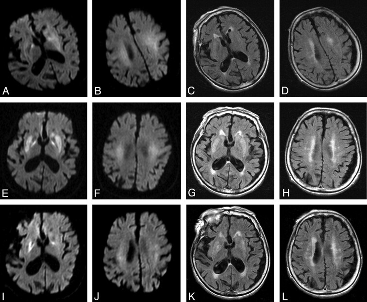
Fig 3.
Follow-up images of hemispheric white matter and basal ganglia lesions on DWI (A, B, E, F, I, J) and FLAIR (C, D, G, H, K, L) (patient 21). The white matter lesions on arrival (A−D) did not disappear by day 2 (E−H) or by 1 week (I−L) after admission. The basal ganglia lesions became more conspicuous by day 2 (E−H) and became slightly less visible after 1 week (I−L).

