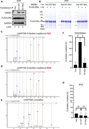
Fig. 3. GREB1 glycosylates ERα on T553 and S554.

(A) HEK293T cells were transfected with FLAG-ERα together with either Vec, GT-WT, or GT-Mut. (B) FLAG pulldown was performed in HEK293T cells described in (A). Total protein eluates were analyzed by Coomassie blue staining. (C to E) O-GlcNAcylation of gel-extracted ERα from the corresponding band from (B) was labeled using BEMAD [β-elimination and Michael addition with dithiothreitol (DTT)] methodology and analyzed by LC-MS/MS. Spectra of ERα tryptic peptide LHAPTSR with modifications at T553 (C) and S554 (D) and with no modification (E) are illustrated. (F) Quantification of the fraction of peptides that were glycosylated at T553 and S554 in the indicated samples. n = 2; *P < 0.05 by one-way ANOVA with Holm-Sidak’s multiple comparisons test. (G) Quantification of the fraction of peptides that were glycosylated at S10 in the indicated samples. n = 2; by one-way ANOVA with Holm-Sidak’s multiple comparisons test.
