. 2021 Mar 17;10:e63387. doi: 10.7554/eLife.63387

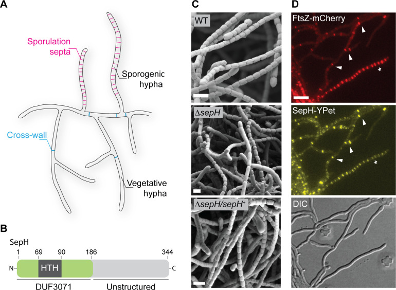
Figure 1. sepH is required for sporulation septation in Streptomyces venezuelae.

(A) Schematic illustrating the multicellular life style of Streptomyces including the two FtsZ-dependent modes of cell division that occur in vegetative and sporogenic hyphae: cross-wall formation and sporulation septation. (B) Schematic of the predicted SepH domain organization including the N-terminal DUF3071 domain containing a helix-turn-helix (HTH) motif and the unstructured C-terminal domain. Numbers indicate corresponding amino acid positions. (C) Cryo-scanning electron micrographs of sporogenic hyphae from wild-type (WT) S. venezuelae, the ΔsepH mutant (SV56), and the complemented mutant strain ΔsepH/sepH+ (MB747). Scale bars: 2 μm. (D) Subcellular co-localization of fluorescently labeled FtsZ (FtsZ-mCherry) and SepH (SepH-YPet) in vegetative and sporulating hyphae. Fluorescent gene fusions were expressed in the WT background (MB751). White arrow heads point at co-localization at cross-walls in vegetative hyphae and the asterisk denotes a sporogenic hypha undergoing sporulation septation. Scale bar: 5 µm.

Figure 1.

Figure 1—figure supplement 1. sepH is a direct target of the transcriptional regulators WhiA and WhiB.

Figure 1—figure supplement 1.

ChIP-seq traces showing the enrichment of the FLAG-tagged developmental regulators WhiA and WhiB at binding sites upstream of sepH (vnz_27360) or their absence in the untagged wild-type (WT) control sample. Source data: Bush et al., 2013.

Figure 1—figure supplement 2. Spore length analysis of wild-type (WT) S. venezuelae, the ΔsepH mutant (SV56), and the complemented mutant strain ΔsepH/sepH+ (MB747).
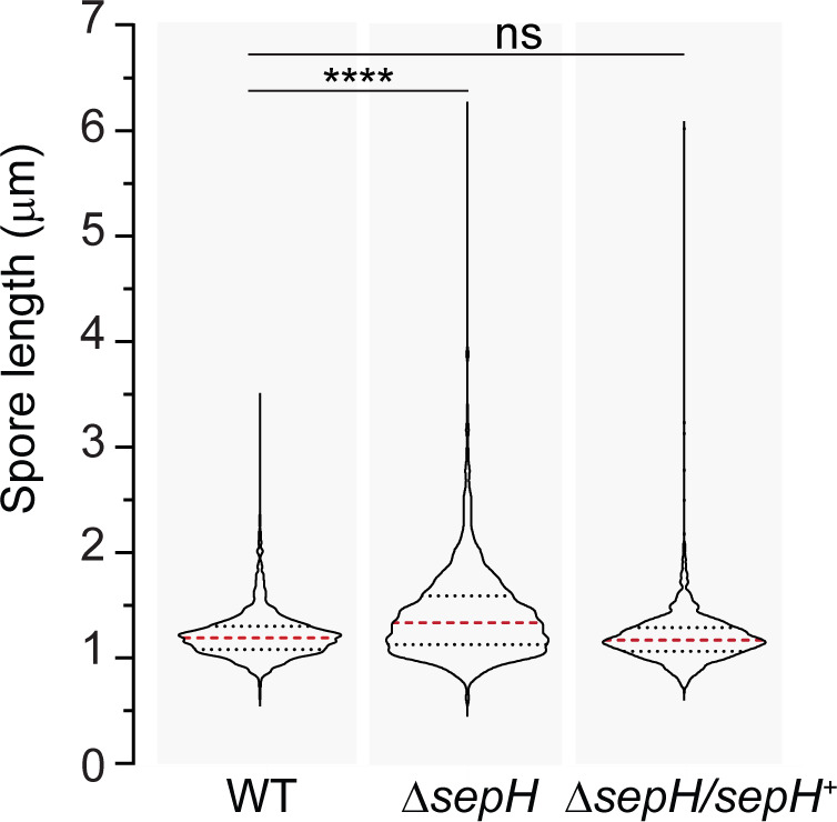
Figure 1—figure supplement 2.

A minimum of 347 spores were quantified for each biological replicate (n = 3) and strain. The dashed red lines indicate the median, and black dotted lines the 25/75th percentiles. Statistical comparisons were made using a one-way ANOVA test followed by a Dunnett’s multiple comparison test comparing the means to the WT mean. ****p<0.0001; ns, not significant.
Figure 1—figure supplement 2—source data 1. Spore size measurement data.

Figure 1—figure supplement 3. Localization and corresponding protein abundance of SepH-YPet in the wild-type (WT) and the ΔftsZ mutant.

Figure 1—figure supplement 3.

(A) Localization pattern of constitutively produced SepH-YPet in the WT (MB858) and in an ΔftsZ mutant strain (MB859). Scale bar: 5 μm. (B) Virtual automated Western blot showing the accumulation of SepH-YPet produced from the constitutive ermE* promoter in MB858, MB859, and an untagged WT control carrying the empty vector (e.v., SS4). YPet fusions were detected with an anti-GFP antibody (1:200). Shown are representative results of duplicate experiments.