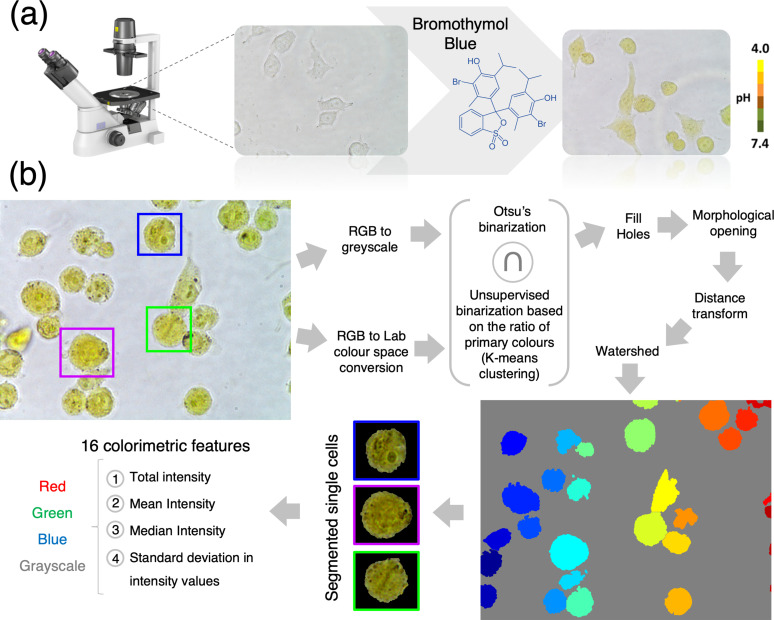
FIG. 1.
Single-cell pH-based imaging, segmentation, and feature extraction workflow. (a) Workflow of the colorimetric pH imaging. Color images are acquired using an optical microscope equipped with a color digital camera. (b) Automated single-cell segmentation pipeline. Raw images are converted to the L*a*b color space and each pixel classified as being a “background pixel” or “cell pixel” using k-means clustering. In parallel, RGB images are converted to the grayscale and thresholded using Otsu's method. A mask is created by selecting only the pixels that were classified as cell pixel by both methods simultaneously. Watershed is used to segment individual cells. Finally, 16 colorimetric features are expected from each cell.

