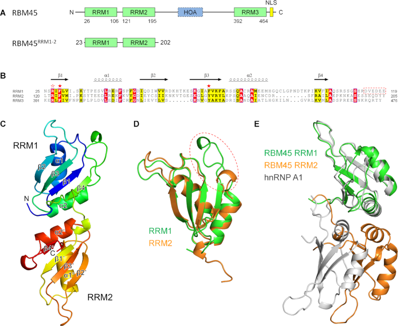
Figure 1.
The structure of RBM45RRM1–2. (A) The domains of human RBM45. (B) Sequence alignment of RRM1, RRM2 and RRM3. The identical residues and conserved residues are highlighted in the red and yellow background, respectively. The two conserved aromatic residues are denoted by red stars, the conserved arginine residues are denoted by a red blank star. (C) The overall structure of RBM45RRM1–2. (D) Superimposition of RRM1 and RRM2. The RRM1 and RRM2 are shown as green and orange cartoons, respectively. The α2−β4 loop is enclosed in a red dashed circle. (E) Superimposition of RBM45RRM1–2 and hnRNP A1. The RRM1 of RBM45 is superposed with the RRM1 of hnRNP A1 (PDB code: 1HA1). The RRM1 and RRM2 of RBM45 are colored green and orange, respectively, the hnRNP A1 is colored gray.

