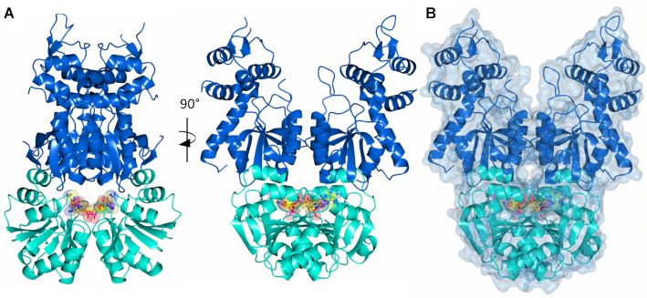
Figure 4.
Structure of Can2 bound to cA4 activator. (A) Two views of the Can2 dimer in cartoon representation. The CARF domain is shown in cyan and the nuclease domain in blue. A molecule of cA4 is bound across the CARF dimer, which is shown as spheres (carbon in yellow, oxygen in red, nitrogen in blue, phosphate in magenta). (B) Surface representation of Can2 dimer with the same colouring as (A).

