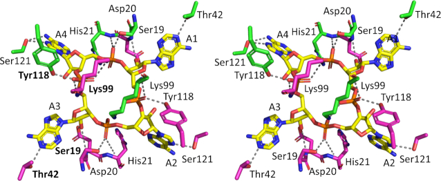
Figure 6.
Structural comparison of cA4 bound to Can2. Divergent stereo representation of cA4 (in stick representation; carbon in yellow, nitrogen in blue, oxygen in red; phosphorus in orange) in complex with the Can2 CARF domain dimer (in stick representation; each monomer in the dimer is coloured green or magenta). Each AMP is numbered (A1–A4). The dotted lines represent hydrogen bond interactions. The residue labels in bold indicate there is a conserved interaction between cA4 in both Can 1 and Can2.

