Figure 4.

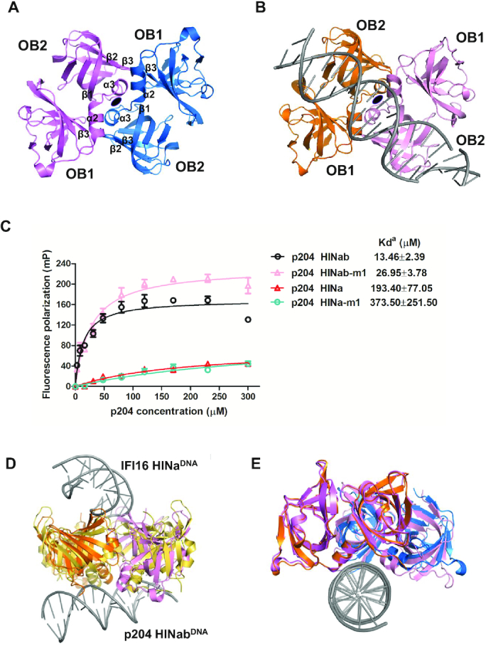
The dimeric interface of p204 HINa domain. (A) HINa dimer in p204 HINa crystal. Two HINa molecules are related by a 2-fold rotational asymmetry and coloured differently. The secondary structure components involved in the dimeric interface were labeled. (B) HINa dimer in p204 HINab:dsDNA complex crystal. (C) FP assay of the dimeric interface of p204 HINab and HINa binding to 30 mer dsDNA. The apparent Kd values (Kda) are shown for p204 HINab, HINa and mutants. (D) Structural comparison of HINa dimer from p204 HINab:DNA and IFI16 HINa:DNA complexes. HINa dimer from IFI16 HINa: DNA complex is shown in yellow orange. HINa dimer from p204 HINab:DNA is shown in orange and pink. (E) Structural comparison of HINa dimer from p204 HINa and HINab:dsDNA complex. HINa dimer from p204 HINa structure is shown in violet and slate. HINa dimer from p204 HINab:dsDNA complex is shown in orange and pink.
