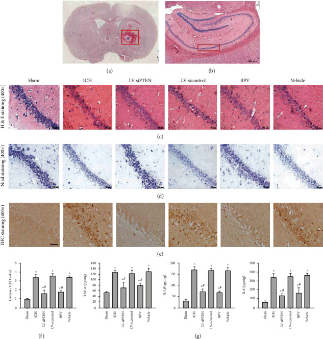
Figure 3.

PTEN knockdown attenuated ICH-induced secondary hippocampal injury. (a–e) Representative images of H&E staining (a, b, c), Nissl staining (d), and caspase-3 IHC staining (e) in the hippocampal CA1 region at 3 d after ICH in rats. Images in (a) and (b) showed the site of brain hemorrhage and the CA1 region of the hippocampus, respectively. Bar, 50 μm. (f) The quantitative analysis of caspase-3 protein normalized to the control group. (g) The impacts of PTEN inhibition on hippocampal neuroinflammation by analyzing the contents of TNF-α, IL-1β, and IL-6 in the ipsilateral hippocampus using ELISA analysis at 3 d postinjury. ∗P < 0.05 compared with the Sham group; #P < 0.05 compared with the ICH group.
