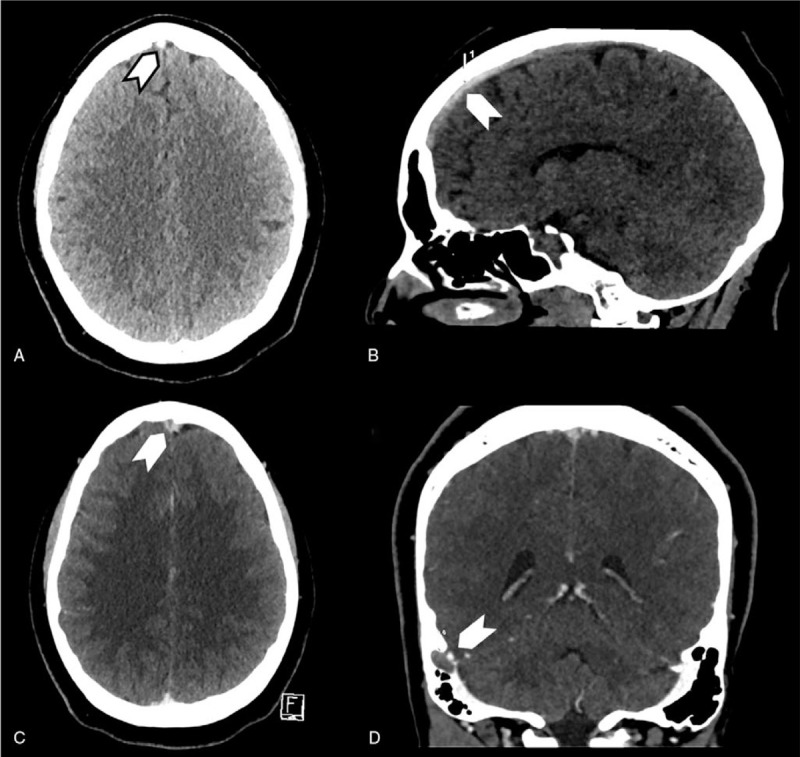
Figure 4.

Patient (case 2) cerebral CT scan showing cerebral venous thrombosis. A (axial view) and B (sagittal view) showing spontaneous density of the anterior part of the superior sagittal sinus (arrows), C (axial view) and D (sagittal view) with contrast showing filling defect in the superior sagittal sinus with classical delta sign (C) and in the right sigmoid sinus (D) (arrows).
