Figure 8.

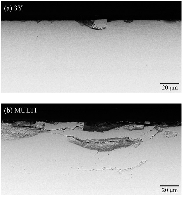
Cross-sectional SEM images of the sub-surface damage introduced in 3Y (a) and MULTI (b) specimens after 500,000 cycles. The sliding direction of the antagonist is from left to right (black arrow).

Cross-sectional SEM images of the sub-surface damage introduced in 3Y (a) and MULTI (b) specimens after 500,000 cycles. The sliding direction of the antagonist is from left to right (black arrow).