
FIGURE 3.
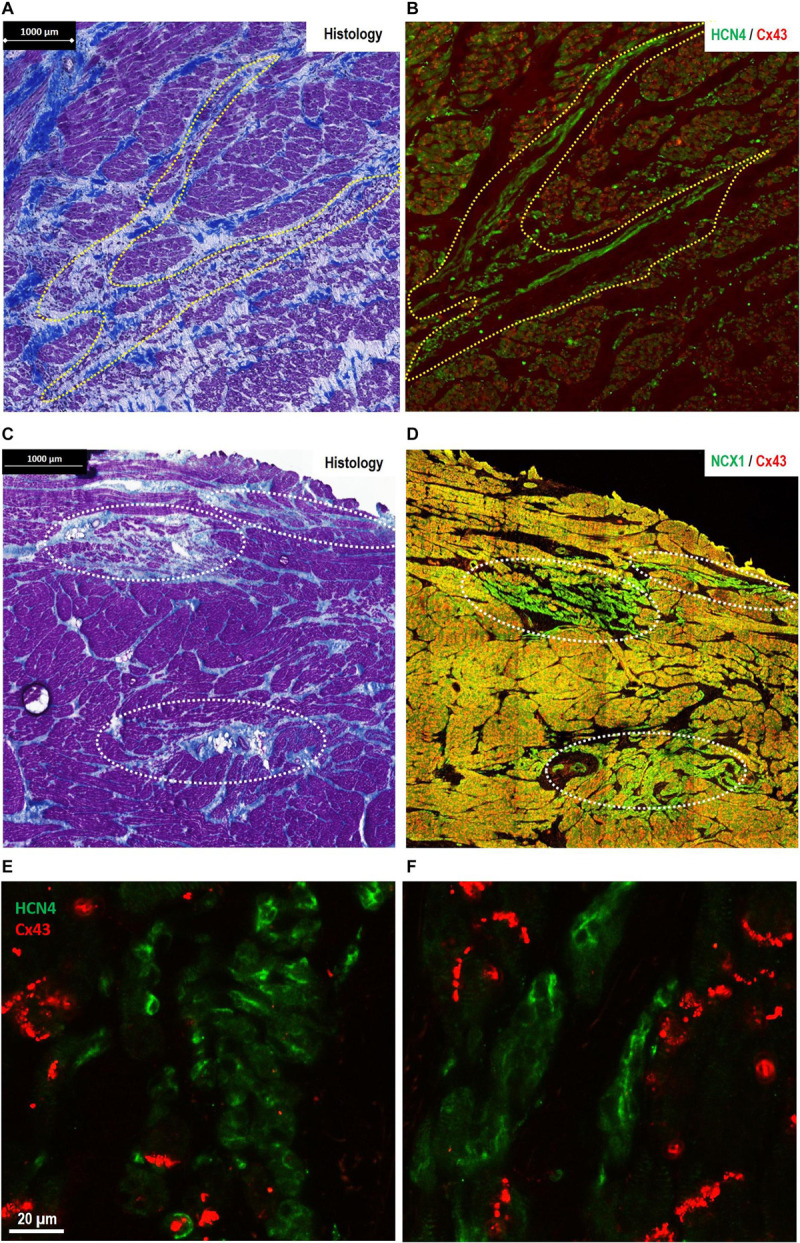
Morphological features of pacemaker/sinus node (SAN)-like tissue in regions other than the primary SAN [where the site of earliest activation (SEA) is detected in healthy hearts]. (A,B) The finger-like projections of pacemaker-like tissue in the paranodal area (PNA)—“witch fingers.” (A) Histology (Masson’s trichrome) section of the PNA from a control goat (outlined in yellow, A). (B) Location of nodal-like cells are confirmed by immunofluorescence labeling of an adjacent section double labeled for Cx43 (red) and HCN4 (green). The nodal-like “witch” fingers in the PNA are predominantly HCN4+/Cx43–. (C,D) Islands of pacemaker-like cells within the subsidiary atrial pacemaker (SAP) regions—“lonely islands.” (C) Masson’s trichrome staining showed that cells with nodal-like phenotype in the SAP region formed well-defined islands in sections taken from the CT. (D) These “lonely islands” are confirmed to have “nodal”-like phenotype by IF labeling of an adjacent section Cx43 (red) and NCX1 (green). The nodal-like island SAP are predominantly NCX1+/Cx43−. Corresponding high-power images of sections stained for HCN4/Cx43 are shown for SAP islands in panel (E) and “witch fingers” in panel (F) demonstrating clusters of HCN4+/Cx43− pacemaker-like cells (green) among the Cx43+ atrial myocytes (red).