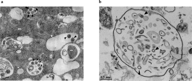
Fig. 2. Ultrastructural visualisation of internalised RSV.
a Transmission electron microscopy of RSV particles within W. magna food vacuoles after 72 h of co-culture. b Purified released extracellular vesicle after 24 h of co-culture containing RSV virions. Virions were randomly selected and measured using ImageJ (marked 3–10). Three morphology categories of RSV were found: spherical (black arrowheads), asymmetric (marked av), and filamentous (marked fv). mitochondria (m), vacuole (v).

