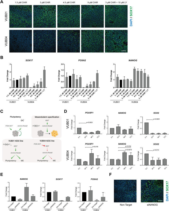
Figure 5.
Stronger activation of WNT signalling improves DE differentiation efficiency of VUB04. (A) Representative immunofluorescent images for SOX17 after 72-h DE differentiation of VUB01 and VUB04 in various differentiation conditions. The scale bar represents 100 μm. (B) Gene expression analysis of SOX17, FOXA2 and NANOG in VUB01 and VUB04 differentiated for 72 h towards DE—standard differentiation condition (3 µM CHIR99021 for the first 24 h) was compared to modified conditions (different concentration of CHIR99021 during the first 24 h or additional incubation with PI3K inhibitor LY294002 for the first 48 h). Data represents three biological replicates. (C) Schematic illustration of the signalling pathways involved in the maintenance of pluripotency (white background) and mesendoderm (ME) specification (grey background). The lower part illustrates the difference in signalling pathway crosstalks between VUB01 and VUB04 hESC lines which influence the differentiation outcome. Created with BioRender. (D) Gene expression dynamics of pluripotency markers in VUB01 and VUB04 during the 72-h DE differentiation. Data represents three biological replicates. The p-values were calculated with unpaired t-test. (E) Comparison of NANOG, SOX17 and FOXA2 expression after 72-h DE differentiation between VUB01 and VUB04 transfected cells. Data represents two biological replicates. (F) Representative images for SOX17 after 72-h DE differentiation of VUB04 transfected with either non-targeting or NANOG-targeting siRNA. The scale bar represents 100 μm.

